【衡道丨笔记】肺罕见肿瘤1例
时间:2023-11-18 21:20:56 热度:37.1℃ 作者:网络
临床病史
-
男,63岁;
-
因痰中带血6月于外院胸部CT发现右肺上叶后段见实性结节,双侧胸膜增厚;
-
我院核医学检查示:右肺中叶体积缩小伴条片状实变,局部糖代谢增高,最大横截面大小约为2.4*1.3cm,毗邻支气管闭塞,右肺门见糖代谢异常增高淋巴结,较大者大小约为0.7*0.6cm;无吸烟史,无唾液腺、头颈部、乳腺肿瘤病史;
-
于我院肺叶切除术+清理分组淋巴结。
镜下
两种形态
第一种:
典型的浸润性肺腺癌形态;
复杂腺体结构、微乳头型,乳头和腺泡,可见STAS。
第二种:
巢状、实性结构伴腔内坏死;
支气管黏膜受累。
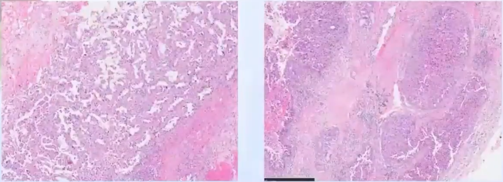
左:第一种形态;右:第二种形态
诊断思路:
-
低分化肺腺癌
-
鳞状细胞癌
-
神经内分泌肿瘤
-
涎腺型肿瘤
-
转移源性……
免疫组化小结:两种形态
第一种:
典型的浸润性肺腺癌形态;
阳性:TTF-1、NapsinA、AR(focal)。
第二种:
巢状、实性结构伴腔内坏死;
阳性:AR、GCDFP-15、Her2、P40(基底层)、TRPS1;
阴性:TTF-1、NapsinA、syn、cga、p63、ER、PR。
分子结果:
FISH:Her-2小簇状扩增(阳性)。
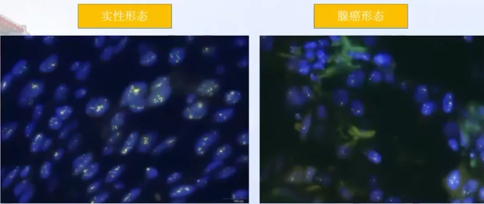
最终病理结果
诊断:
恶性肿瘤,可见两种形态,一种为浸润性肺腺癌,Ⅲ级,复杂腺体结构为主(约60%),并见微乳头成分 (约20%) 和腺泡成分;一种为实体型低分化癌,沿支气管生长,结合形态及免疫组化结果,符合原发肺的涎腺型导管癌。
40基因结果:ERBB2、TP53突变。
FISH:Her-2扩增。
知识回顾
-
肺涎腺型肿瘤是起源于支气管涎腺的一组肿瘤
-
与头颈部涎腺肿瘤为同一类组织起源
-
头颈涎腺肿瘤在肺内均可发生!
-
原发性肺涎腺型导管癌极为罕见
-
自2014年以来开始有个例报道,至今仅10例
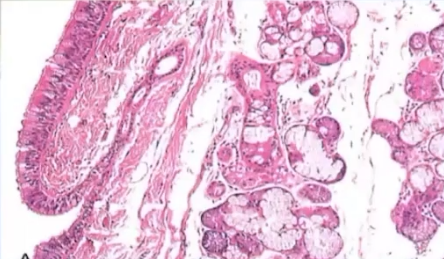
最新肺WHO-涎腺型肿瘤(salivary gland-typetumors):
多形性腺瘤(pleomorphic adenoma);
腺样囊性癌(adenoid cysticcarcinoma);
上皮-肌上皮癌(epithelium-myoepithelial carcinoma);
黏液表皮样癌(mucoepidermoid carcinoma);
玻璃样变透明细胞癌(hyalinizing clear cell carcinoma);
肌上皮瘤和肌上皮癌(myoepithelioma and myoepithelial carcinoma).
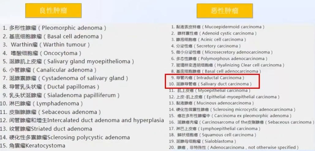
涎腺导管癌
-
1968年被首次报道,罕见;
-
最常发生于腮腺,其次为颌下腺和小涎腺;
-
2005年WHO定义为“类似于高级别乳腺导管癌的侵袭性腺癌”;
-
病理学形态与乳腺浸润性导管癌极为相似;
-
高度侵袭性,易血道转移及淋巴结转移(肺54%);
-
手术切除+术后放化疗;
-
25-90% Her-2过表达或扩增,与预后差相关;
-
64-77% AR过表达,治疗有效。
涎腺导管内瘤
-
1983年被首次命名,罕见;
-
最常发生于腮腺;
-
“低级别涎腺导管癌”、“低度恶性筛状囊腺癌”、“涎腺导管原位癌”等命名被先后使用;
-
2017年WHO正式命名为“涎腺导管内癌”;
-
类似乳腺导管原位癌或ADH的导管内增生性病变;
-
巢周肌上皮证实原位癌的本质;
-
可以伴微小浸润,也分低级别和高级别;
-
生物学行为较为惰性、复发转移少;
-
形态及分子分型:RET重排、P53突变等。
因具有不同的的临床病理特征和生物学行为,认为二者是两个不同的肿瘤实体!
但通常发生于肺时,可以两种成分共同存在。
文献复习-原发肺的涎腺型导管癌
首篇(2014)

-
A 55-year-old woman,No cigarette smoking history.
-
No past cancer history of the salivary gland, head and neck, or the breast.
-
CT:a solid mass of size 3*3*3cm in the central part of left upper lobe.
-
A lobectomy was performed.
-
After surgery,adjuvant radiotherapy was given.
-
The patient died of respiratory failure and distant brain metastasis 19 months after initial diagnosis.
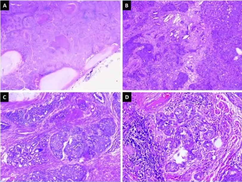
(A)Intraductal component
(B)Irregular invasive solid nests
(C)Lobular cancerization
(D)Lobular cancerization
原位癌(小叶癌化)+浸润瘤
诊断方向:
阴性:
-
腺:TTF-1,Napsin-A
-
鳞:P40 p63
-
神经内分泌:Syn cga
-
MEC and ACC:Mucicarmine CD117
阳性:
-
AR:strong nuclear staining
-
GCDFP-15 :focally positive
-
HER2/neu gene amplification(lHC+FISH)
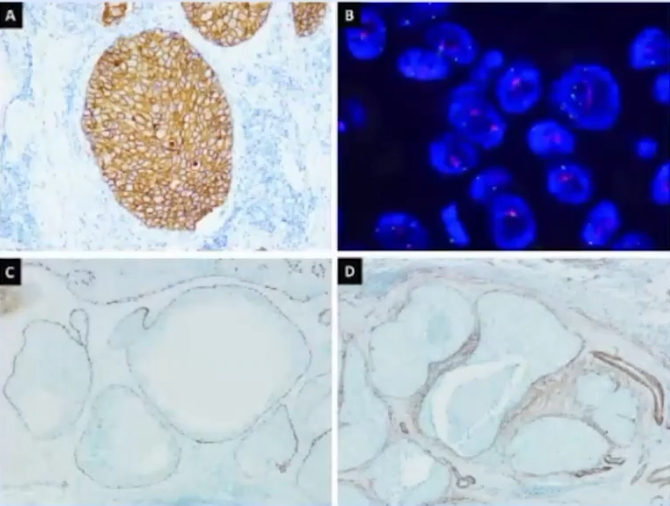
第二篇(2016)
-
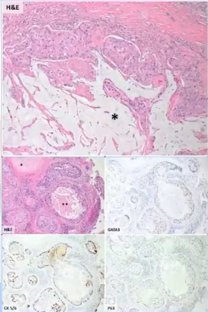
a case of a 73-year-old woman,active smoker,the right upper lobe.
-
an exceedingly rare subtype of an already rare malignant salivary-type neoplasm.
-
bears a rare BRAF G464V mutation.
-
distinctive histologic and immunophenotypic features resembling DCIS of the breast.
-
no history of malignancy.
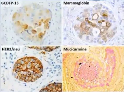
鉴别诊断
-
High-grade mucoepidermoid carcinoma
-
mixed mucinous/non-mucinous AC
-
Metastatic
免疫组化
-
focal AR stains
-
TTF1, napsin A,ER,and PR are negative (not shown)
分子
-
exon 11 of BRAF,c.1391GNT (p.G464V),22%
第三篇(2020)

-
An 82-year-old male had been followed-up for 2 years for smoking-related interstitial pneumonia.
-
Gross examination revealed a gray-white tumor meas-uring 15 mm adjacent to the pleura.
-
Right lower lobe lobectomy was performed.
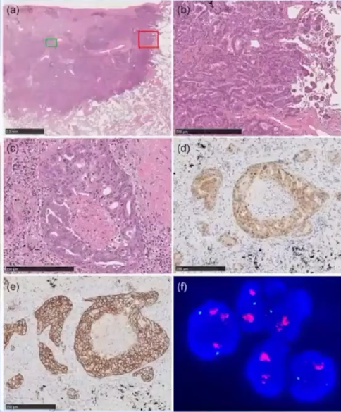
(a)The tumor is located mainly in the subpleural lung with spread into the surrounding alveoli
(b)Closer views of the green and red squares are shown in(b)and (c)
(d)The tumor cells are positive for AR
(e)The cellular membranes of the tumor cells are strongly positive for HER2
提出疑问
-
a novel category,cribriform predominant adenocarcinoma,which is frequently negative for TTF-1, has been proposed.
-
several studies have also reported that a small proportion of lung adenocarcinomas show HER2/neu amplification and/or AR expression.
第四篇(2022年,上海肺科医院)
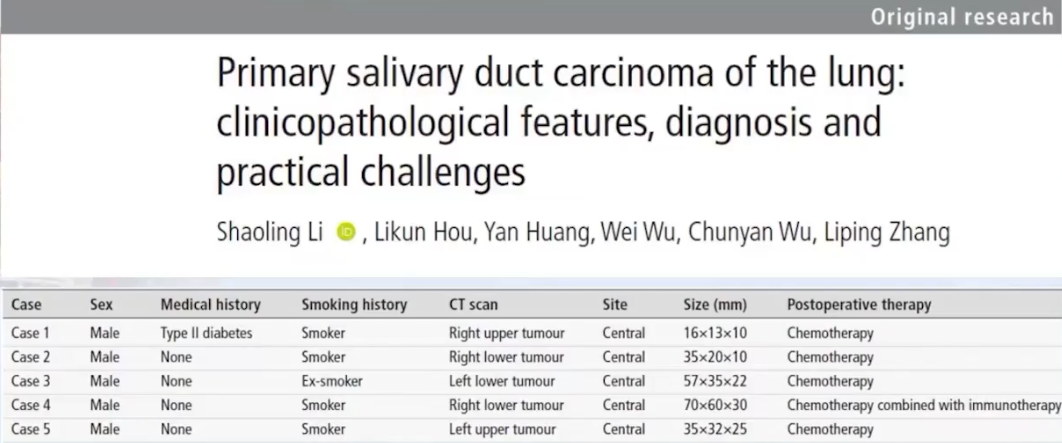
均为男性,均有吸烟史,均为中央型
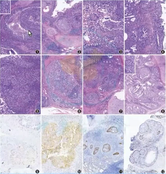
形态特点
导管内生长(原位,提示原发)+浸润性生长;
实性巢状伴粉刺样坏死;
可有乳头状生长方式;
支气管黏膜下腺体呈现“小叶癌化”特征;
可形成筛状结构,类似“罗马桥”结构。
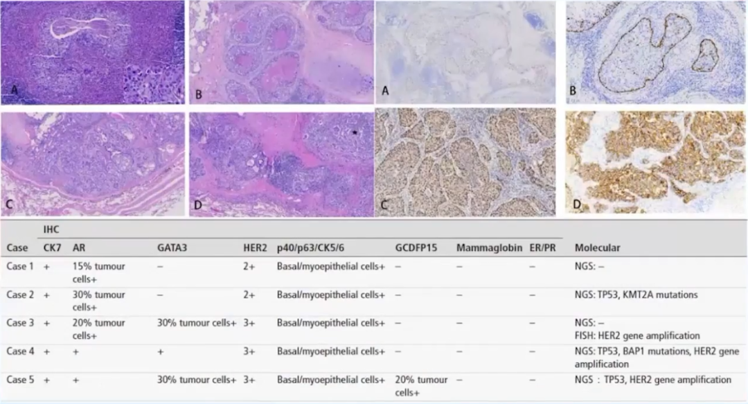
免疫组化
AR 10-15%+
Her2 2+
P63 CK5/6示基底细胞/肌上皮
TTF1,napsin A,ER,PR,P40,GATA3,GCDFP15 均阴性
PSA P504S均阴性
二代测序
例1未见基因异常,例2发生P53和KMT2A突变
此篇文献中总结的鉴别诊断
-
低分化肺腺癌;
-
原发鳞状细胞癌;
-
涎腺分泌型癌:排列多样,腔隙常见嗜酸性分泌物,核分裂和粉刺样坏死不多见,胞质内常见分泌空泡,共表达S100和mammaglobin,ETV6-NTRK3基因融合;
-
乳腺浸润性导管癌肺转移:病史影像+肺内导管内癌(双层结构,提示原发);
-
头颈部涎腺型导管癌肺转移:只能通过病史影像;
-
20%的头颈部涎腺型导管癌可表达PSA,因此男性患者需排除前列腺癌肺转移,导管内癌双层结构支持肺原发。
第五篇(2023年,上海肺科医院)
小结
发生部位:多为中央型:年龄、性别、有无吸烟目前无统计学差异;
镜下形态:导管内癌(原位癌)+浸润癌,两种形态混杂存在,或以一种形态为主;
导管内癌:类似乳腺导管原位癌,可见粉刺样坏死,可有乳头结构;
浸润性瘤:可实性、小巢状、微乳头状浸润性生长;
原位特征:“小叶癌化”或Paget样累及支气管黏膜;
免疫组化:
阳性:AR、GCDFP15、GATA3、TRPS-1、HER-2、基底细胞;
阴性:腺、鳞、神经内分泌表型。
鉴别诊断:腺、鳞、神经内分泌、其他涎腺型肿瘤(包括分泌性癌);
分子:Her-2扩增。
治疗及预后
-
手术+清扫淋巴结+术后辅助放化疗免疫治疗;
-
AR阳性患者可通过抗雄激素疗法获益;
-
有文献报道,个例患者可以从曲妥珠单抗中获益,获得长期生存;
-
预后:个例患者因脑转移死亡,OS为19个月。


